Contrast-enhanced mammography (CEM) combines mammographic imaging of the breast with intravenous administration of contrast media. In addition to showing anatomical structures, it can highlight areas with increased contrast-media uptake. Find out more about how the examination is performed, how our Accutron® CT-D Vision can be used for the procedure, and what experience from clinical practice has shown.
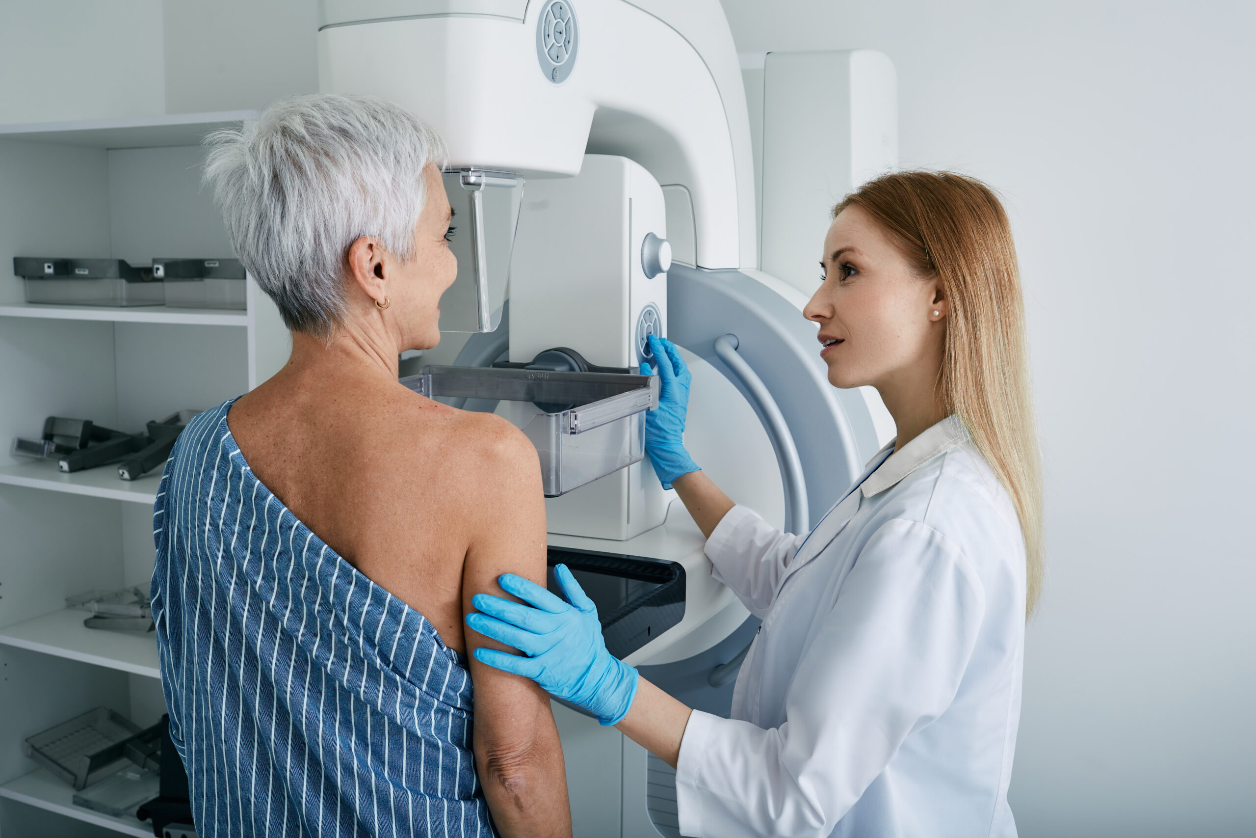
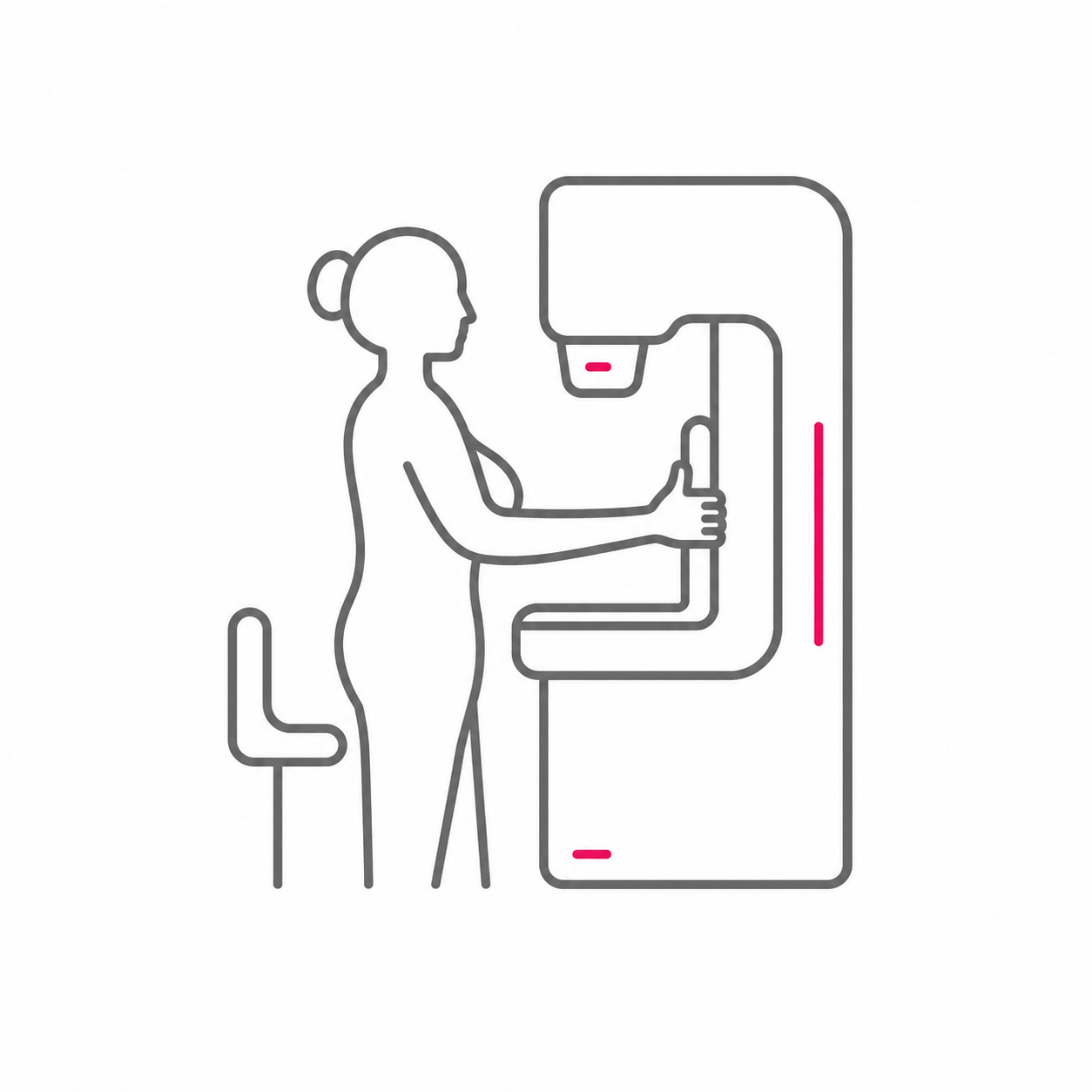
Contrast-enhanced mammography is an X-ray examination of the breast. It is used to further investigate abnormal findings following conventional mammography or ultrasound. Contrast-enhanced mammography combines digital mammography with intravenous administration of an iodine-based contrast media.
During the examination, both low- and high-energy images are acquired. The system then uses both sets of images to create additional recombined images. The low-energy images show the anatomical and morphological structures of the breast, as in a conventional mammogram. By contrast, iodine is much more strongly contrasted in the high-energy images, while normal breast tissue looks almost the same in both. The computer can subtract the low-energy images from the high-energy images, leaving an image showing almost nothing but the contrast media.
So in summary: CEM produces two complementary types of images: a conventional mammogram showing the structures of the breast, and an image highlighting areas where the contrast media has accumulated.
Dr Sabine Ohlmeyer, Head of Gynaecological Radiology and Multimodal Breast Imaging at the Institute of Radiology, University Hospital Erlangen
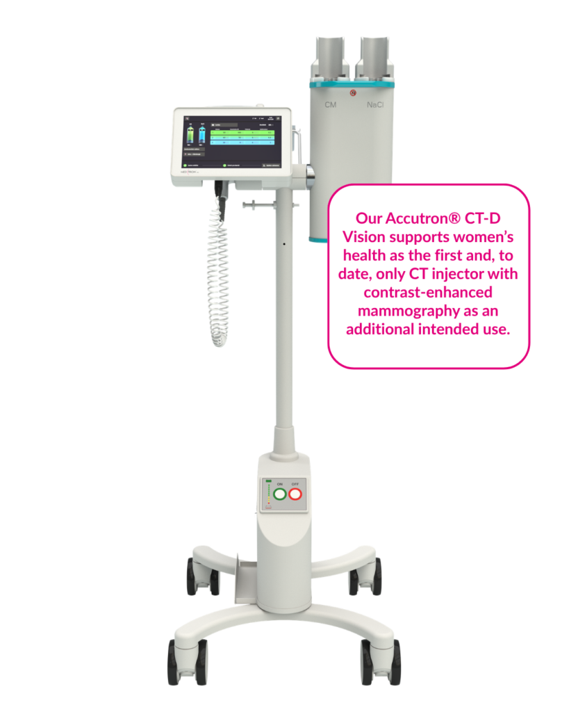
The Accutron® CT-D Vision from MEDTRON delivers contrast media continuously and precisely during CEM imaging. This enables uniform contrast enhancement of the tumour tissue. The injector can also be used to inject saline afterwards, helping to move the contrast media more quickly onwards from the arm veins. As a result, the contrast media spends less time in the veins, which can help reduce irritation of the vein walls. The injector is wireless and mobile, so it can be easily moved to the patient and conveniently used as part of the examination.
A contrast-enhanced mammogram usually takes about 15 to 30 minutes. This includes preparation, placing an intravenous line, administering the contrast media and taking the mammographic images. Imaging usually begins a few minutes after the injection starts and is performed within a planned time window. The precise time required may vary depending on the examination protocol and the work-flow at the centre.
CEM can be used in a range of situations, for example to further assess abnormal or unclear findings, evaluate the extent of confirmed breast cancer, support preoperative planning, and monitor the response to neoadjuvant treatment during or after it has been administered. CEM can also provide an additional or alternative imaging option when breast MRI cannot be performed or is not available. The choice between the two imaging methods depends on the individual clinical situation.

Usually, less than one minute. The precise time required depends on the contrast-media volume and the chosen flow rate. Imaging then begins within the planned time window specified in the protocol.
CEM uses an iodine-based X-ray contrast media, which is administered intravenously. The contrast media used for CEM is the same type as that used for other X-ray-based examinations, such as computed tomography. Before the examination, standard medical procedures are followed to identify any contraindications and individual risk factors, such as allergies.
CEM is generally no more painful than conventional mammography. In both procedures, brief compression of the breast can cause a sensation of pressure or discomfort. CEM also involves insertion of an intravenous line and administration of the contrast media. This may briefly cause a feeling of warmth or a metallic taste.
Sie werden zu einer externen Website weitergeleitet:
Für die Inhalte der externen Website ist ausschließlich deren Betreiber verantwortlich.