Discover the fascinating world of contrast media injection in our MEDTRON Knowledge Hub. Immerse yourself in comprehensive information about the different types of contrast media, injection techniques and safety guidelines. Our platform offers multimedia content to expand your expertise and give you a deeper understanding of this important technology in medical imaging. Whether you are an experienced professional or simply curious, you will find valuable resources to deepen your knowledge of contrast media injection and stay up to date.
CONTRAST MEDIA PROTOCOL
RECOMMENDATIONS
FROM HEAD TO FOOT
What is the optimal amount of contrast medium for a CT scan of the skull? What flow rate is suitable for abdominal or thoracic examinations? In daily practice, many parameters must be determined quickly and reliably.
Together with Alex Riemer, we have created a comprehensive document with practical contrast medium protocol recommendations for CT examinations. The clear tables provide structured guidance on important parameters such as contrast agent volume, concentration, injection rate and examination phases – broken down by different examination regions from head to toe.
The document serves as practical support for radiologists and MTRs to efficiently plan contrast agent protocols and support standardised CT procedures.
The ‘Golden Rules of Contrast Media Administration’ – concise, understandable and straight from the everyday life of a CT expert. Written by Alex Riemer and provided exclusively by MEDTRON AG, they offer valuable guidance for safe and efficient contrast media administration. With many years of practical experience and a trained eye for the crucial details, Alex Riemer summarises the most important basic principles of modern contrast agent administration. Whether for newcomers or experienced users, this article offers valuable insights for everyday radiology practice.
#1 Duration of injection for arterial CT examinations: alignment with the duration of the scan
For CT examinations targeting only arteries (e.g. CT angiography, arterial chest CT), the acquisition scan duration is the main factor taken into consideration when configuring the injection protocol.
This means: The injection duration must be proportionate to the acquisition duration.
If this is not taken into account, the injection duration may be too long or too short, resulting either in an unnecessarily high dose of contrast media or in suboptimal contrast enhancement.
#2 Calculation of the contrast media quantity for arterial examinations
The contrast media quantity for an arterial examination can be calculated using the following formula:
CM quantity [ml] = (scan time [s] + delayaBT [s]) x flow [ml/s]
DelayaBT = delay after bolus tracking – the time delay between reaching the trigger threshold and the start of the spiral scan
#3 Influence of body weight on contrast and flow in CT examinations
If the contrast media protocol is not adapted to the patient’s body weight, the following will apply:
The higher the patient’s body weight, the poorer the contrast enhancement.
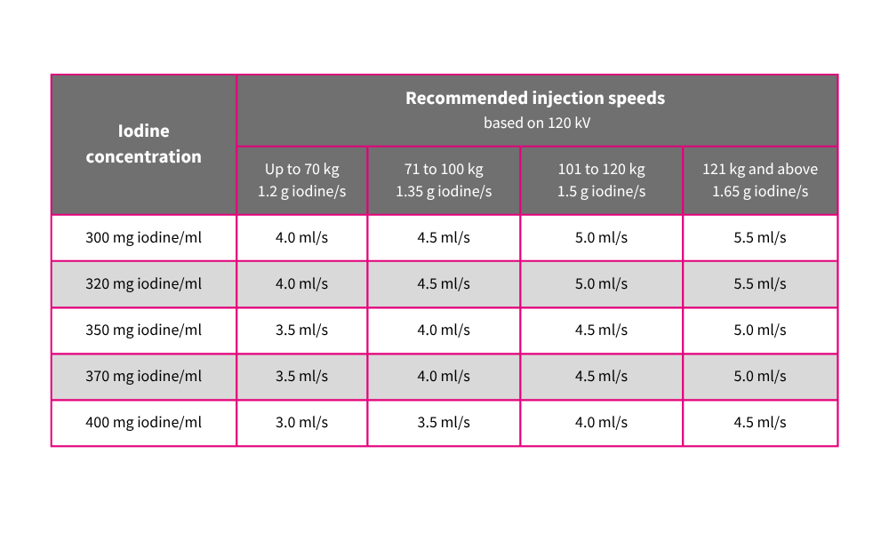
For arterial examinations:
The higher the patient’s body weight, the higher the flow rate should be (Figure 1).
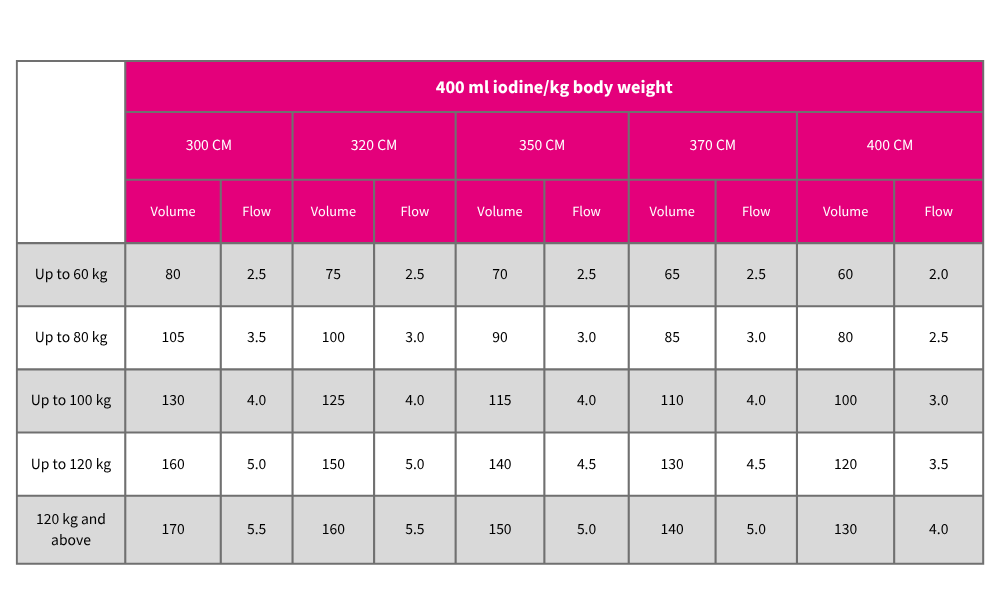
For abdominal parenchymal examinations (e.g. liver):
The higher the patient’s body weight, the higher the contrast media volume should be (360-500 mg iodine/kg body weight) (Figure 2).
In order to achieve effective contrast enhancement irrespective of body weight in the arterial phase of an abdominal CT examination as well, the flow rate should be adapted in addition to the CM volume.
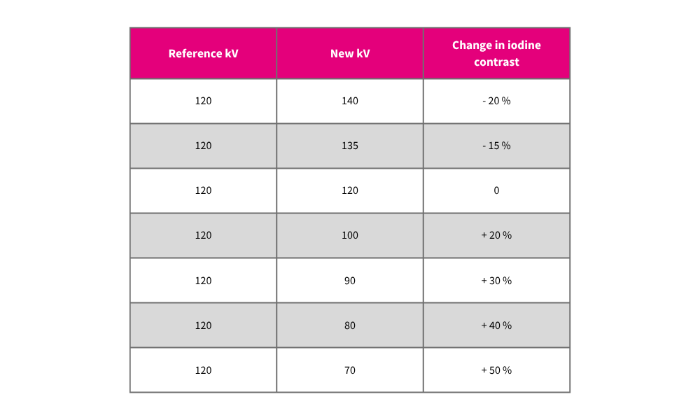
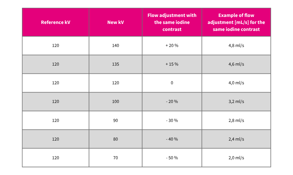
#4 Tube voltage and iodine contrast: the correlation in the CT protocol
Tube voltage has a considerable influence on iodine contrast (Figure 3).
If the contrast media protocol is not adapted, the following will apply:
The higher the tube voltage, the poorer the iodine contrast.
The lower the tube voltage, the better the iodine contrast.
For arterial examinations (e.g. CTA) combined with poor injection conditions, a low tube voltage can help to achieve adequate arterial contrast enhancement in spite of a low flow rate.
#5 Target value for vascular contrast in CT angiography: at least 250 HU
For CT angiography, vascular contrast of at least 250 HU should be achieved.
#6 Optimal Hounsfield units for diagnostic liver CT: more than 100 HU
For an effective diagnostic liver CT scan, the parenchymal contrast of the liver should be greater than 100 Hounsfield units (HU).
#7 The rule for contrast-enhanced CT: "CM is the way forward!"
For contrast-enhanced CT examinations, this rule should be followed wherever possible:
“CM is the way forward!”
CT examinations should be performed with a sufficient quantity of CM to ensure an accurate diagnosis.
If the contrast media volume and/or flow rate is too low, it may not be possible to establish a diagnosis.
#8 Elimination of iodinated contrast media with normal kidney function
With normal kidney function, intravenously administered iodinated contrast media are 50% eliminated after around 2 hours, 75% eliminated after 4 hours and almost completely eliminated after 24 hours.
#9 Bolus tracking positioning: Ascending or descending aorta?
Whether the bolus tracking/test bolus ROI is positioned in the ascending aorta or descending aorta has no relevant influence on the examination quality or the iodine contrast, as the blood flow velocity in the thoracic aorta is typically between 0.72 m/s and 1.2 m/s.
#10 Higher iodine concentration for improved contrast with low flow
The higher the iodine concentration of the contrast media, the better the contrast will be, even with low contrast media quantities and flow rates.
About the Author
Alex Riemer is an expert in computed tomography, an enthusiastic MTR, a passionate trainer and speaker, and a successful author of specialist books.